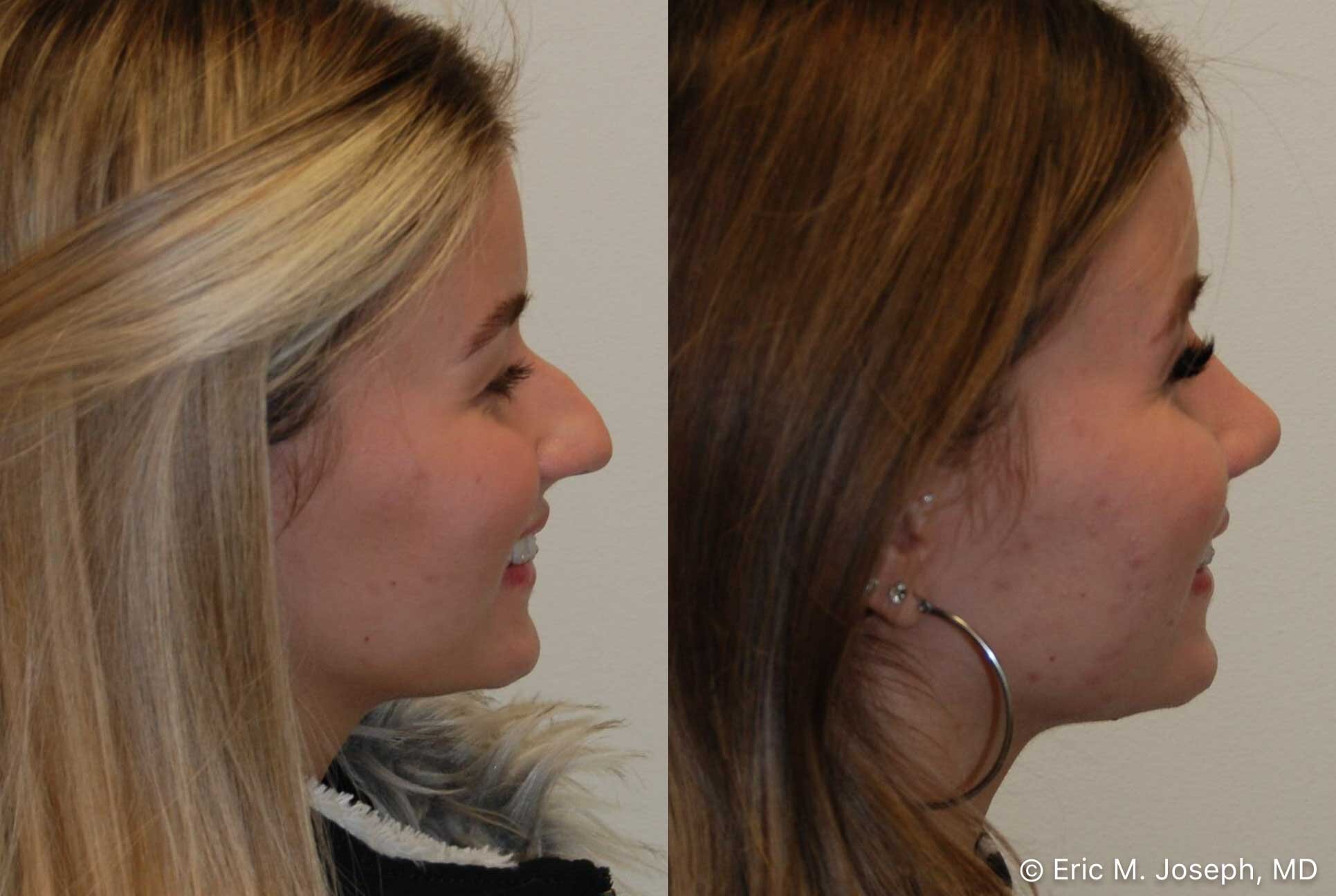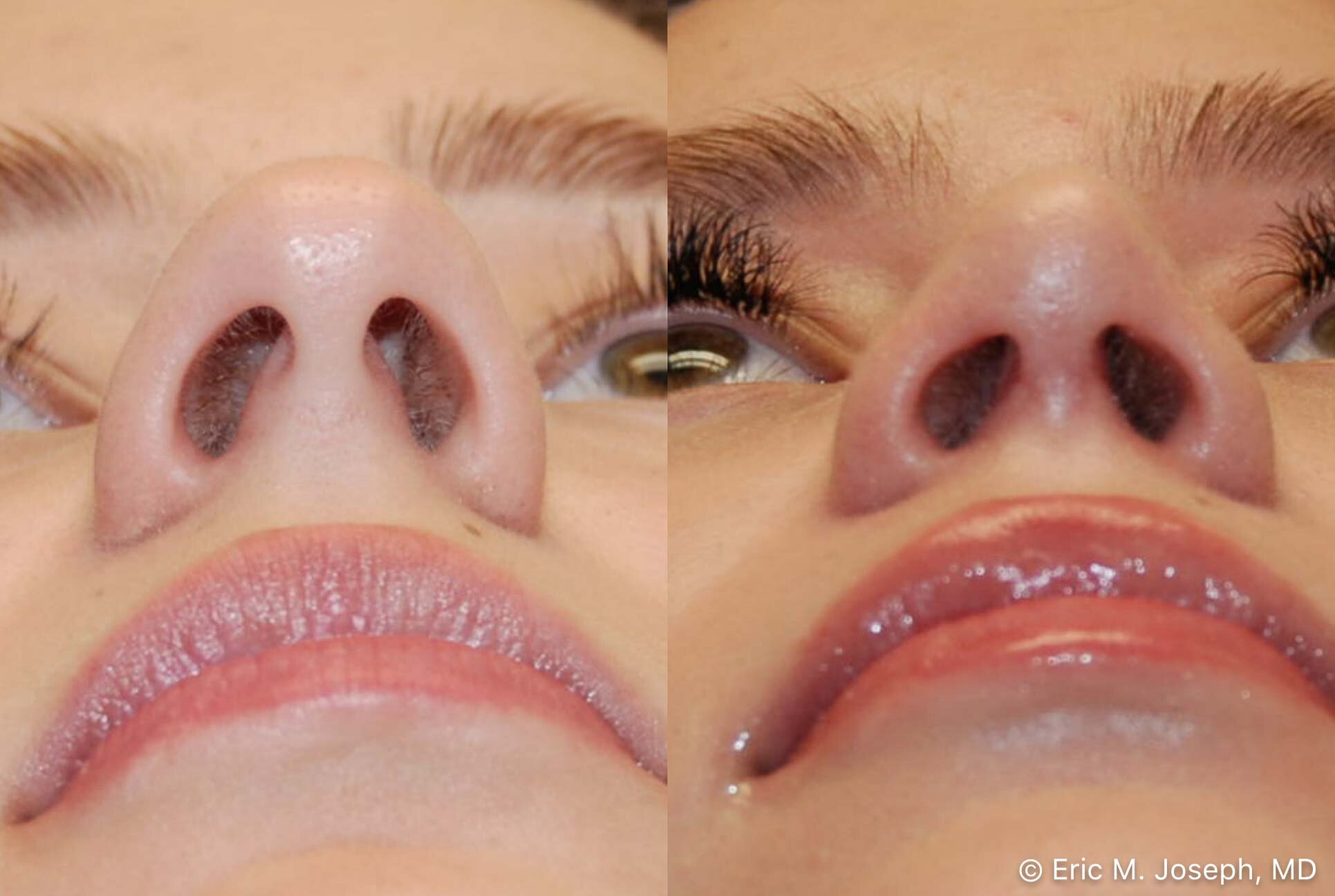


Rhinoplasty #193
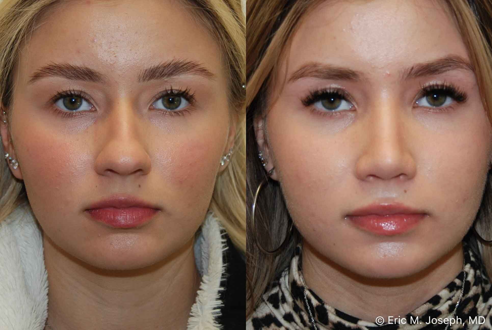
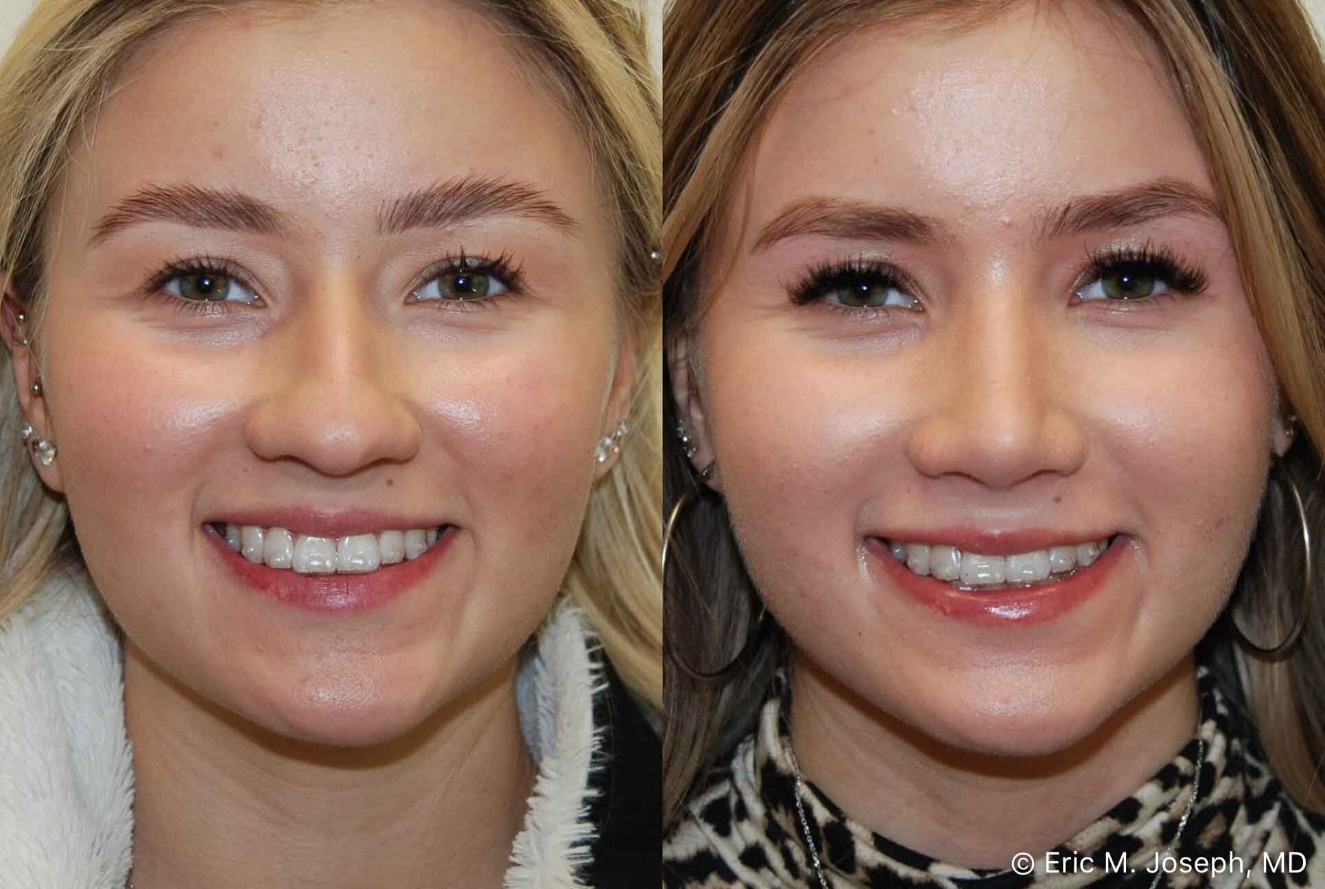
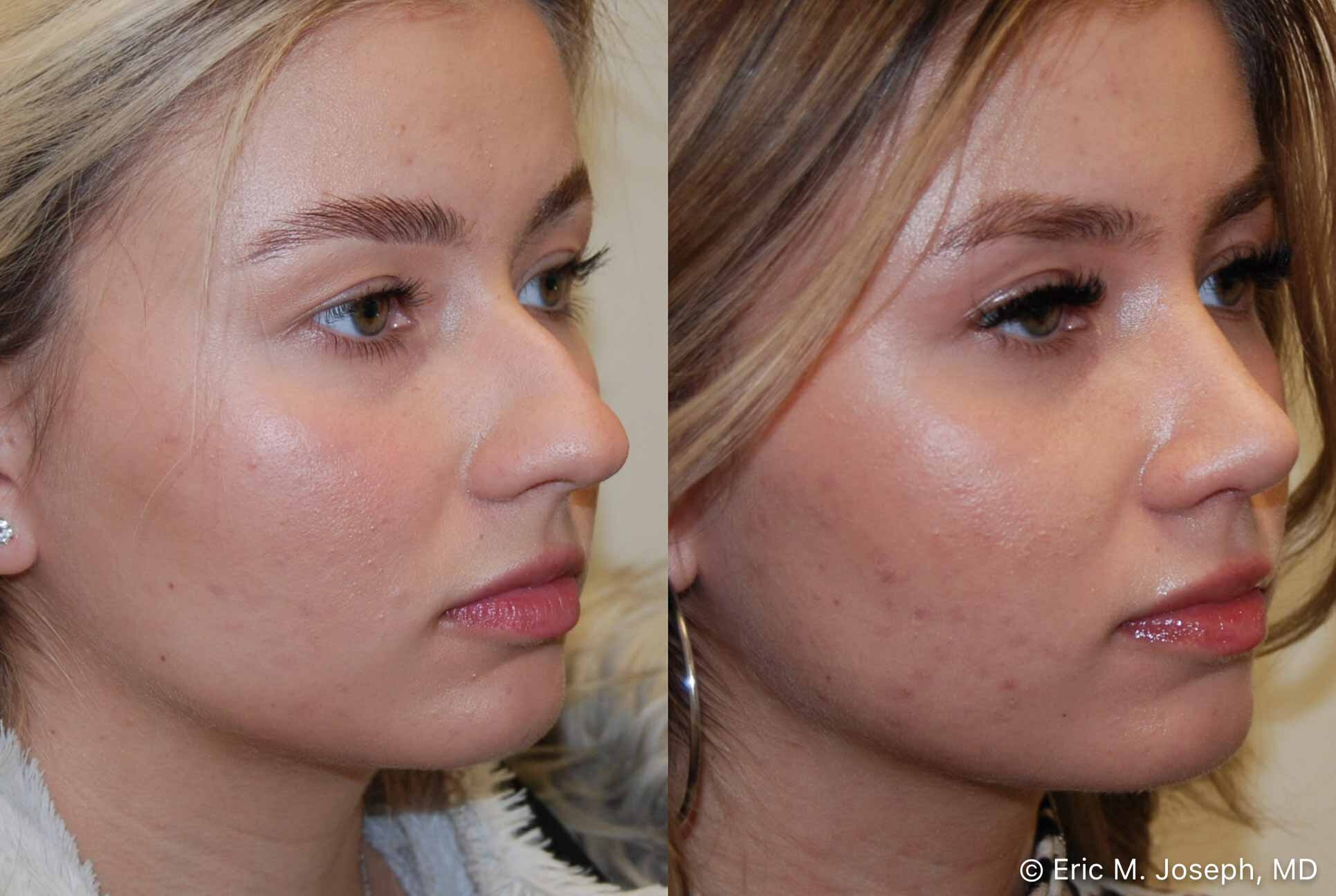
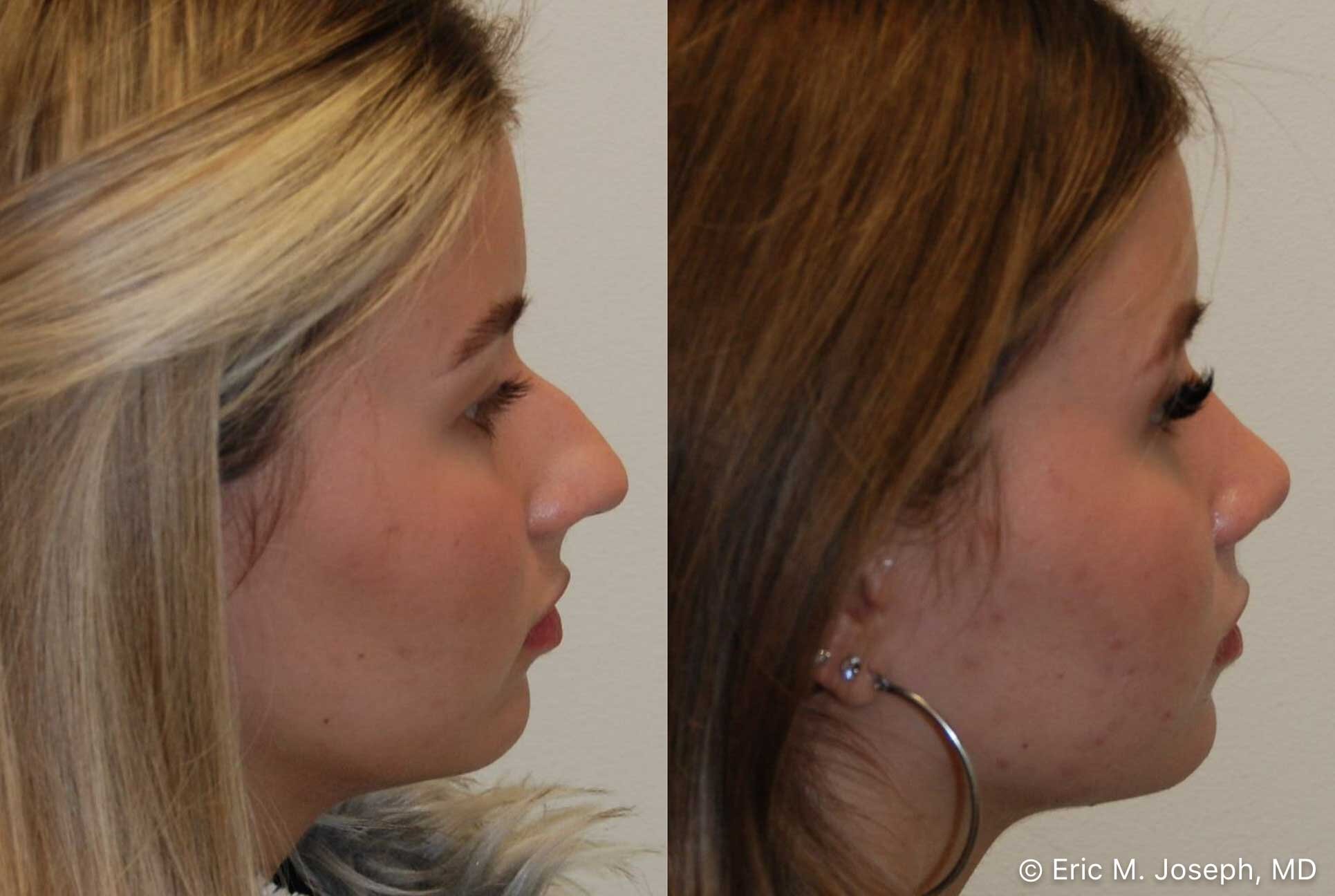
Our beautiful teenage patient is seen 7 months following cosmetic rhinoplasty surgery. She was bothered by the excessive width of her nose, and her profile bump. Her surgery consisted of narrowing of her bridge, tip, and nostrils, with hump removal, and tip projection. Before surgery, her tip was a bit flat and underprojected. We used an extended shield graft to add definition and projection to her nasal tip.
Some patients that have visited us for rhinoplasty consultations have been surprised that other surgeons have neither examined nor touched their noses. We feel the examination of one's nose is critical for determining one's candidacy for rhinoplasty surgery. What follows describes what a nasal examination would be like if you're considering nose job surgery:
While sitting at your side, your face is gently positioned using both hands, so you're looking straight ahead. I use both index fingers to feel the sidewalls of your nose starting from the glabella down to the tip while noting the thickness and mobility of your nasal skin, and the firmness of your underlying cartilaginous and bony nasal framework.If we see you have light-colored, thin nasal skin, and firm adherent tip cartilages, you may be told that you may be more prone to encounter visible irregularities after rhinoplasty compared to others with thicker nasal skin. We assess the patency of each nasal airway by gently occluding each nostril with the pad of the thumb, without distorting the nasal appearance, and requesting you to breathe normally through your nose several times. We use direct head lighting to illuminate your nasal airway and visualize your internal nasal anatomy. We also lift the tip with one thumb, and view the position of the caudal septum, and the size and color of the inferior turbinates. The undersurface of your lower lateral tip cartilages are examined while palpating with the thumb and index finger. The thumb lifts your nostril margin, and the index finger pushes the dome of the tip down to see the internal configuration of your tip, and assess the quality and size of your alar cartilages. Next we examine your nasal profile. The size and shape of your nose, and its proportion with your upper lip, lower face and chin are noted. Gentle palpation begins at your nasal bones and sequentially moves down the bony-cartilaginous junction to the tip of your nose. Your nasal tip is then palpated from side to side and from top to bottom with two fingers. The strength of your lower lateral cartilages and the thickness and adherence of your tip skin are assessed. The length, position and lateral mobility of the bottom of your nasal septum is palpated with the thumb and index finger, and the spatial relationship between your medial crura, caudal septum and alar rim is also noted. If you have breathing issues that need to be addressed, we may decongest your turbinates with nasal spray, and examine both your internal and external nasal valves. A nasal speculum is used to spread your nostrils gently open for further visibility inside your nose. We sometimes use a 3 millimeter flexible fiberoptic endoscope to examine structures further inside your nose to ensure you do not have nasal polyps or enlarged adenoids. We find the examination is critical for determining whether we can help someone with rhinoplasty surgery. For this reason, we will not agree to operate on anyone until we are able to perform a thorough nasal examination.


